CHROMOSOME ANALYSIS AND CLASSIFICATION
For this purpose cytogenetic studies or chromosome cultures have to be performed on rapidly dividing somatic cells from the metaphase stage of mitosis. The commonly used cells are peripheral lymphocytes, fibroblasts from skin biopsies, cells from bone marrow biopsies or cells from any other tissue biopsy. Foetal cells from an amniocentesis, cells from chorionic villi extracted by a chorionic villi sampling manouvre or foetal cells like blood cells from cordocentesis can be used. Liver cells and skin cells taken from tissue biopsies using foetoscopy procedures, or blood cells using the same procedure can also be used as culture material.
Briefly the procedure of chromosome culture from peripheral lymphocytes consists of firstly collecting a sample of intravenous peripheral blood using sterile equipment and procedures into a sterile heparinised container. The sample is next treated with growth promoting culture media containing essential ingredients including balanced salt solutions and a substance called Phytohaemagglutinin (PHA) which is a mitogen that stimulates cell division. Fig. 15. These steps are carried out in a sterile laminar flow cupboard, using sterile techniques and sterile containers. The resulting mixture is next incubated at 37 degrees Celsius for three days. Shortly before the end of this period of time, the culture is treated with a spindle poison called colchicine which inhibits the spindle. The medium is next replaced with hypotonic saline to swell the chromosomes and next fixed using a mixture of alcohol and acetic acid.
Fig 15
The resulting solution is now spread out on slides, dried and stained with specialized stains which give each homologous pair of chromosomes a unique banding pattern. When trypsin and Giemsa stain is used for the purpose, the pattern is referred to as G banding. Fig 16. Other staining methods are the use of fluorescent dyes and examination under UV light.
The slides are then examined and spreads (groups) of 46 are analysed. The good spreads are selected and photographed using a photomicroscope. The film is now developed, the photographs printed and the individual chromosomes are next cut out and arranged in pairs in decreasing order of size, and numbered from 1 to 22 with the sex chromosomes separate. This photographic representation of the entire somatic chromosome complement is referred to as a KARYOTYPE. Fig. 17. A normal female karyotype is denoted as 46, XX and that of a male as 46, XY. The main bands and the sub bands are numbered beginning from the centromere and moving towards the tips of the chromosome and the missing or exchanged or rearranged bands are denoted in the final formula. Fig. 16.
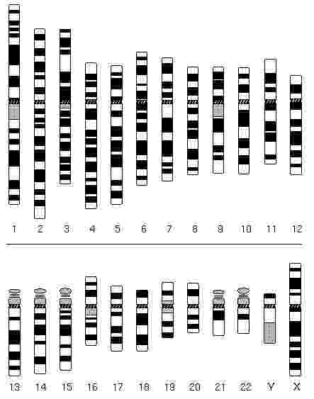
Fig 16
Fig 17
High resolution banding, is another technique where chromosomes are arrested in a less coiled, longer form. This method is helpful in detecting small changes or losses in chromosome aberrations.